Median raphe cysts represent infrequent benign lesions that develop due to abnormalities during embryonic formation of the urethral folds. Epidermal inclusion cysts arising along this anatomical line are particularly rare and can be mistaken for other penile swellings. They often appear as small, firm white or yellow swelling causing discomfort or tenderness. It can be congenital or acquired by epithelial cell implantation into the skin. It frequently occurs in young men often appearing in the second to third decade, although they are present from childhood. We describe a case involving a 24 months-old male who presented with a painless cystic lesion along the median raphe of the penis since birth. The swelling was managed by complete surgical removal, and histological evaluation confirmed the diagnosis of an epidermal inclusion cyst. Recognition of this condition is important to ensure correct diagnosis and appropriate treatment.
This is an Open Access article, distributed under the terms of the Creative Commons Attribution 4.0 International License (http://creativecommons.org/licenses/by/4.0/), which permits unrestricted use, distribution and reproduction in any medium or format, provided the original work is properly cited.
Epidermal Inclusion Cyst, Median Raphe Cyst, Penile Lesion, Congenital Anomaly
1. Introduction
The median raphe extends along the ventral surface of the penis from the urethral opening to the perineum and corresponds to the line formed during fusion of the urethral folds in embryonic life
[1]
Amaranathan A, Sinhasan SP, Dasiah SD. Median raphe cysts of the prepucial skin, with triple histological linings: a case report and review of the literature. J Clin Diagn Res. 2013; 7(7): 1466–68.
. Lesions arising along this tract, referred to as median raphe cysts, are uncommon and encompass different histological types such as urethral cyst, epidermal inclusion cyst, and dermoid cysts
[4]
Nagore E, Sánchez-Motilla JM, Febrer MI, Aliaga A. Median raphe cysts of the penis: a report of five cases. Pediatric Dermatology. 1998; 15: 191–93.
. Although usually asymptomatic, it may lead to cosmetic concerns or patient anxiety due to their location. Awareness of such lesions helps clinicians differentiate them from other penile pathologies and avoid unnecessary interventions. While distinguishing benign cyst may have limited clinical significance, since local incision is the preferred definitive treatment in most cases, improved recognition is important to avoid confusion in premalignant conditions
[12]
López-Ríos F, Rodríguez-Peralto JL, Castaño E, Benito A. Squamous cell carcinoma arising in a cutaneous epidermal cyst: case report and literature review. Am J Dermatopathol. 1999; 21: 174–77.
Chiu MY, Ho ST. Squamous ce Nishida H, Kashima K, Daa T, Yada N, Arakane M, Matsuda K, Kato A, Shimizu F, Yokoyama S. J Cutan Pathol. 2012 Aug; 39(8): 808-10.
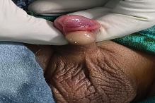
A 24-months-old male reported in surgical outpatient department with a painless swelling about 1.5 cm in diameter over the ventral aspect of the penis. The swelling had been present since birth, but was noticed recently by his parents due to slight increase in its size. The lesion had shown gradual enlargement over time but was not associated with pain, discharge, urinary complaints or any features of inflammation. There was no relevant history of trauma, infection, or prior procedures. On the basis of above clinical findings a provisional diagnosis of median raphe cysts of penis was made with differential diagnosis of urethral cyst, dermoid cyst and epidermal inclusion cyst.
2.1. Clinical Examination
l examination revealed a single, well-demarcated, dome-shaped swelling measuring about 1.5 × 1 cm, located along the median raphe of the penile shaft. The lesion was soft, non-tender and mobile, with normal overlying skin, not adherent to skin. No regional lymph node enlargement was observed (Figure 1).
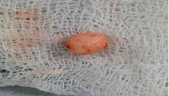
Figure 3. Excised specimen measuring approximately 1.5 × 1 cm with smooth outer surface.
Cut section
On cut section - a thicken walled cystic space seen, filled with dirty color amorphous necrotic material.
3.2. Microscopic
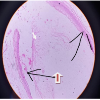
On microscopic examination - cyst wall is thickened with fibro collagenous stroma. Underlying surface is lined by stratified squamous epithelium with a distinct granular layer. Cyst is filled with keratin debris and flecks (Figure 4).
(Figure 4). There were no features suggestive of malignancy
[12]
López-Ríos F, Rodríguez-Peralto JL, Castaño E, Benito A. Squamous cell carcinoma arising in a cutaneous epidermal cyst: case report and literature review. Am J Dermatopathol. 1999; 21: 174–77.
Recovery was uneventful. At follow-up after six months, no recurrence was detected, and the parents were satisfied with the outcome and longer follow-up is planned.
4. Discussion
Median raphe cysts are thought to originate from developmental disturbances during formation of the male urethra, possibly due to incomplete fusion or entrapment of epithelial elements
[1]
Amaranathan A, Sinhasan SP, Dasiah SD. Median raphe cysts of the prepucial skin, with triple histological linings: a case report and review of the literature. J Clin Diagn Res. 2013; 7(7): 1466–68.
. Basically it is of two types, urethroid and epidermoid. Based on whether the epithelium is of urethra or epidermal tissue. The epidermal subtype is relatively rare and is often identified in adolescents or young adults
[3]
Unsal A, Cimentepe E, Saglam R. Penile epidermoid cyst in an elderly patient. Int Urol Nephrol. 2002; 34(2): 229–30.
. Typically, such lesions are slow-growing and symptom-free but may occasionally become noticeable due to enlargement or cosmetic concerns.
Conditions that may resemble median raphe cysts include dermoid cysts, epidermoid cyst of glans penis and mucous cyst of penis
[14]
Lezcano C, Chaux A, Velazquez EF, Cubilla AL. Semin Diagn Pathol. 2015 May; 32(3): 245-8. ll carcinoma arising from an epidermal cyst. Hong Kong Med J. 2007; 13: 482–84.
[15]
Khana S. Epidermoid cyst of the glans penis. Eur Urol. 1991; 19: 176–77.
such as glomus tumors, and infective lesions.These cysts can also arise as a complication of penile girth enhancement surgery.
[10]
Park HJ, Park NC, Park SW, Jern TK, Choi KU. Penile epidermal inclusion cyst: a late complication of penile girth enhancement surgery. J Sex Med. 2008; 5(9): 2238–40.
Careful clinical assessment usually provides sufficient guidance; however, imaging techniques like ultrasonography may be considered when deeper extension or urethral involvement is suspected.
Definitive identification relies on histopathological evaluation
[2]
Okeke LI. Epidermal inclusion cyst as a rare complication of neonatal male circumcision: a case report. J Med Case Rep. 2009; 3: 7321.
. Though immunohistochemistry is highly useful for urothelial origin of Median raphe cyst.
[8]
Dini M, Baroni G, Colafranceschi M. Median raphe cyst of the penis: a report of two cases with immunohistochemical investigation. The American Journal of Dermatopathology. 2001; 23(4): 320–24.
. As there were no skin adnexa, especially pilosebaceous structures within the cyst wall and hair follicles, dermoid cyst is ruled out.
Steatocystoma simlex of the penis is also ruled out, as there was no eosinophilic inner lining of cyst wall with empty lumen having no recognizable cyst content.
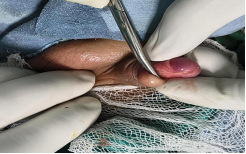
Surgical removal of the lesion in total is regarded as definitive management and generally results in excellent outcomes
[2]
Okeke LI. Epidermal inclusion cyst as a rare complication of neonatal male circumcision: a case report. J Med Case Rep. 2009; 3: 7321.
. There is no documented evidence suggesting malignant change in such lesions. Malignant transformation is extremely rare but has been reported in isolated cases
[12]
López-Ríos F, Rodríguez-Peralto JL, Castaño E, Benito A. Squamous cell carcinoma arising in a cutaneous epidermal cyst: case report and literature review. Am J Dermatopathol. 1999; 21: 174–77.
Chiu MY, Ho ST. Squamous ce Nishida H, Kashima K, Daa T, Yada N, Arakane M, Matsuda K, Kato A, Shimizu F, Yokoyama S. J Cutan Pathol. 2012 Aug; 39(8): 808-10.
. Sometimes a huge penile mass is benign and noncancerous if it turns out to be an epidermal inclusion cyst.
[11]
Kaviani A, Hosseini J, Vazirnia AR. A huge penile mass which turned out to be an epidermoid inclusion cyst. Urol J. 2009; 6: 135–37.
[11]
. They rarely turns in to malignancy. They should be considered when evaluating cystic lesions of the penile shaft. Confirmation through histopathology and management by complete excision leads to favorable cosmetic and clinical results with a very low likelihood of recurrence
[6]
Saini P, Mansoor MN, Jalali S, Sharma A. Penile epidermal inclusion cyst. Indian J. Pediatr. 2010; 77(7): 815–16.
1. The present case highlights a classical clinical presentation with clear surgical and histopathological correlation, which adds to the limited pool of documented cases and reinforces awareness among clinicians. The epidermal variant is particularly rare.
2. Histopathological analysis is essential for confirmation.
3. Surgical excision offers definitive treatment with minimal recurrence. median raphe cyst are uncommon developmental lesions of the genital midline
Author Contributions
Nilay Kumar: Conceptualization, Writing – original draft
The authors declare that there are no conflicts of interest.
References
[1]
Amaranathan A, Sinhasan SP, Dasiah SD. Median raphe cysts of the prepucial skin, with triple histological linings: a case report and review of the literature. J Clin Diagn Res. 2013; 7(7): 1466–68.
Dini M, Baroni G, Colafranceschi M. Median raphe cyst of the penis: a report of two cases with immunohistochemical investigation. The American Journal of Dermatopathology. 2001; 23(4): 320–24.
Park HJ, Park NC, Park SW, Jern TK, Choi KU. Penile epidermal inclusion cyst: a late complication of penile girth enhancement surgery. J Sex Med. 2008; 5(9): 2238–40.
Kaviani A, Hosseini J, Vazirnia AR. A huge penile mass which turned out to be an epidermoid inclusion cyst. Urol J. 2009; 6: 135–37.
[12]
López-Ríos F, Rodríguez-Peralto JL, Castaño E, Benito A. Squamous cell carcinoma arising in a cutaneous epidermal cyst: case report and literature review. Am J Dermatopathol. 1999; 21: 174–77.
Chiu MY, Ho ST. Squamous ce Nishida H, Kashima K, Daa T, Yada N, Arakane M, Matsuda K, Kato A, Shimizu F, Yokoyama S. J Cutan Pathol. 2012 Aug; 39(8): 808-10.
Lezcano C, Chaux A, Velazquez EF, Cubilla AL. Semin Diagn Pathol. 2015 May; 32(3): 245-8. ll carcinoma arising from an epidermal cyst. Hong Kong Med J. 2007; 13: 482–84.
[15]
Khana S. Epidermoid cyst of the glans penis. Eur Urol. 1991; 19: 176–77.
Kumar, N., Mandal, A., Singh, S. K., Khadatkar, S. V. (2026). Epidermal Inclusion Cyst of the Median Raphe of the Penis: A Case Report. International Journal of Clinical Urology, 10(1), 92-95. https://doi.org/10.11648/j.ijcu.20261001.26
Kumar, N.; Mandal, A.; Singh, S. K.; Khadatkar, S. V. Epidermal Inclusion Cyst of the Median Raphe of the Penis: A Case Report. Int. J. Clin. Urol.2026, 10(1), 92-95. doi: 10.11648/j.ijcu.20261001.26
Kumar N, Mandal A, Singh SK, Khadatkar SV. Epidermal Inclusion Cyst of the Median Raphe of the Penis: A Case Report. Int J Clin Urol. 2026;10(1):92-95. doi: 10.11648/j.ijcu.20261001.26
@article{10.11648/j.ijcu.20261001.26,
  author = {Nilay Kumar and Aninda Mandal and Sandip Kumar Singh and Saurabh Vijayrao Khadatkar},
  title = {Epidermal Inclusion Cyst of the Median Raphe of the Penis: A Case Report},
  journal = {International Journal of Clinical Urology},
  volume = {10},
  number = {1},
  pages = {92-95},
  doi = {10.11648/j.ijcu.20261001.26},
  url = {https://doi.org/10.11648/j.ijcu.20261001.26},
  eprint = {https://article.sciencepublishinggroup.com/pdf/10.11648.j.ijcu.20261001.26},
  abstract = {Median raphe cysts represent infrequent benign lesions that develop due to abnormalities during embryonic formation of the urethral folds. Epidermal inclusion cysts arising along this anatomical line are particularly rare and can be mistaken for other penile swellings. They often appear as small, firm white or yellow swelling causing discomfort or tenderness. It can be congenital or acquired by epithelial cell implantation into the skin. It frequently occurs in young men often appearing in the second to third decade, although they are present from childhood. We describe a case involving a 24 months-old male who presented with a painless cystic lesion along the median raphe of the penis since birth. The swelling was managed by complete surgical removal, and histological evaluation confirmed the diagnosis of an epidermal inclusion cyst. Recognition of this condition is important to ensure correct diagnosis and appropriate treatment.},
 year = {2026}
}
TY  - JOUR
T1  - Epidermal Inclusion Cyst of the Median Raphe of the Penis: A Case Report
AU  - Nilay Kumar
AU  - Aninda Mandal
AU  - Sandip Kumar Singh
AU  - Saurabh Vijayrao Khadatkar
Y1  - 2026/05/26
PY  - 2026
N1  - https://doi.org/10.11648/j.ijcu.20261001.26
DO  - 10.11648/j.ijcu.20261001.26
T2  - International Journal of Clinical Urology
JF  - International Journal of Clinical Urology
JO  - International Journal of Clinical Urology
SP  - 92
EP  - 95
PB  - Science Publishing Group
SN  - 2640-1355
UR  - https://doi.org/10.11648/j.ijcu.20261001.26
AB  - Median raphe cysts represent infrequent benign lesions that develop due to abnormalities during embryonic formation of the urethral folds. Epidermal inclusion cysts arising along this anatomical line are particularly rare and can be mistaken for other penile swellings. They often appear as small, firm white or yellow swelling causing discomfort or tenderness. It can be congenital or acquired by epithelial cell implantation into the skin. It frequently occurs in young men often appearing in the second to third decade, although they are present from childhood. We describe a case involving a 24 months-old male who presented with a painless cystic lesion along the median raphe of the penis since birth. The swelling was managed by complete surgical removal, and histological evaluation confirmed the diagnosis of an epidermal inclusion cyst. Recognition of this condition is important to ensure correct diagnosis and appropriate treatment.
VL  - 10
IS  - 1
ER  -
Kumar, N., Mandal, A., Singh, S. K., Khadatkar, S. V. (2026). Epidermal Inclusion Cyst of the Median Raphe of the Penis: A Case Report. International Journal of Clinical Urology, 10(1), 92-95. https://doi.org/10.11648/j.ijcu.20261001.26
Kumar, N.; Mandal, A.; Singh, S. K.; Khadatkar, S. V. Epidermal Inclusion Cyst of the Median Raphe of the Penis: A Case Report. Int. J. Clin. Urol.2026, 10(1), 92-95. doi: 10.11648/j.ijcu.20261001.26
Kumar N, Mandal A, Singh SK, Khadatkar SV. Epidermal Inclusion Cyst of the Median Raphe of the Penis: A Case Report. Int J Clin Urol. 2026;10(1):92-95. doi: 10.11648/j.ijcu.20261001.26
@article{10.11648/j.ijcu.20261001.26,
  author = {Nilay Kumar and Aninda Mandal and Sandip Kumar Singh and Saurabh Vijayrao Khadatkar},
  title = {Epidermal Inclusion Cyst of the Median Raphe of the Penis: A Case Report},
  journal = {International Journal of Clinical Urology},
  volume = {10},
  number = {1},
  pages = {92-95},
  doi = {10.11648/j.ijcu.20261001.26},
  url = {https://doi.org/10.11648/j.ijcu.20261001.26},
  eprint = {https://article.sciencepublishinggroup.com/pdf/10.11648.j.ijcu.20261001.26},
  abstract = {Median raphe cysts represent infrequent benign lesions that develop due to abnormalities during embryonic formation of the urethral folds. Epidermal inclusion cysts arising along this anatomical line are particularly rare and can be mistaken for other penile swellings. They often appear as small, firm white or yellow swelling causing discomfort or tenderness. It can be congenital or acquired by epithelial cell implantation into the skin. It frequently occurs in young men often appearing in the second to third decade, although they are present from childhood. We describe a case involving a 24 months-old male who presented with a painless cystic lesion along the median raphe of the penis since birth. The swelling was managed by complete surgical removal, and histological evaluation confirmed the diagnosis of an epidermal inclusion cyst. Recognition of this condition is important to ensure correct diagnosis and appropriate treatment.},
 year = {2026}
}
TY  - JOUR
T1  - Epidermal Inclusion Cyst of the Median Raphe of the Penis: A Case Report
AU  - Nilay Kumar
AU  - Aninda Mandal
AU  - Sandip Kumar Singh
AU  - Saurabh Vijayrao Khadatkar
Y1  - 2026/05/26
PY  - 2026
N1  - https://doi.org/10.11648/j.ijcu.20261001.26
DO  - 10.11648/j.ijcu.20261001.26
T2  - International Journal of Clinical Urology
JF  - International Journal of Clinical Urology
JO  - International Journal of Clinical Urology
SP  - 92
EP  - 95
PB  - Science Publishing Group
SN  - 2640-1355
UR  - https://doi.org/10.11648/j.ijcu.20261001.26
AB  - Median raphe cysts represent infrequent benign lesions that develop due to abnormalities during embryonic formation of the urethral folds. Epidermal inclusion cysts arising along this anatomical line are particularly rare and can be mistaken for other penile swellings. They often appear as small, firm white or yellow swelling causing discomfort or tenderness. It can be congenital or acquired by epithelial cell implantation into the skin. It frequently occurs in young men often appearing in the second to third decade, although they are present from childhood. We describe a case involving a 24 months-old male who presented with a painless cystic lesion along the median raphe of the penis since birth. The swelling was managed by complete surgical removal, and histological evaluation confirmed the diagnosis of an epidermal inclusion cyst. Recognition of this condition is important to ensure correct diagnosis and appropriate treatment.
VL  - 10
IS  - 1
ER  -





 Share This Article
 Twitter
 Linked In
 Facebook
 Copy
Copy Download